Moles come in different shapes, sizes and colors. Some are flat and brown, while others are raised, pink or close to your natural skin tone. Most are harmless, but knowing what is normal for your skin can make it easier to notice a new or changing spot.
In this guide, we explain the main types of skin moles, what they may look like, how to check your skin and when to ask a dermatologist for advice.
What Is a Mole?
A mole, also called a nevus, is a skin growth that develops when pigment-producing cells called melanocytes grow in a cluster. The plural of nevus is nevi.
Moles may be present at birth or appear later in life, especially during childhood and early adulthood. Their appearance can vary depending on a person’s skin tone and the type of mole.
A common mole may be:
- Flat or raised
- Round or oval
- Pink, tan, brown, black or similar to the surrounding skin
- Smooth or slightly textured
- Even in color, with a clearly defined border
These features are common, but they are not a guarantee that a mole is harmless. A dermatologist can assess a mole that is new, changing or otherwise concerning.
Learn more about common benign moles in the Skinive Dermatology Atlas.
Types of Skin Moles
There are several ways to describe moles. Some terms refer to when a mole appeared, others describe how it looks or where its cells are located in the skin. These categories can overlap: for example, a mole can be both acquired and compound.
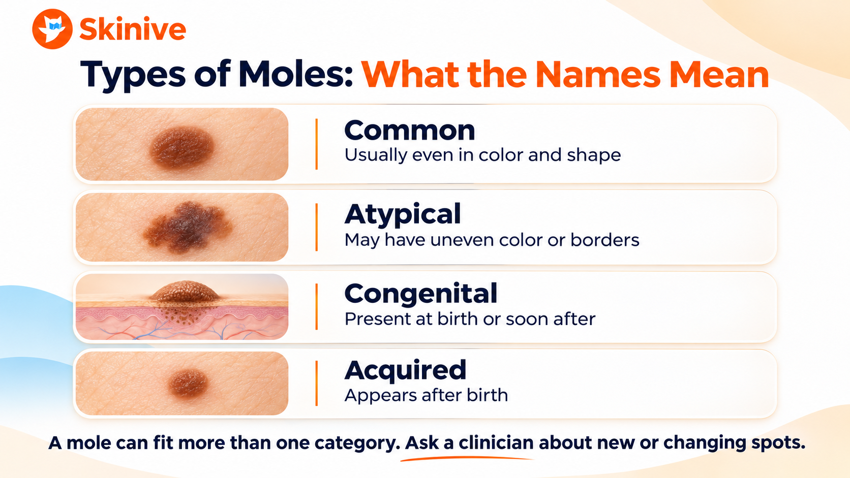
Common Moles
Common moles are usually small, round or oval, with a fairly even color and a distinct border. They can be flat or raised and may appear almost anywhere on the body.
Most common moles are benign, meaning they are not cancerous. However, a mole that changes or begins to look noticeably different from your other moles should be examined.
Atypical Moles: Dysplastic Nevi
An atypical mole, sometimes called a dysplastic nevus, may look different from a common mole. It can have an irregular shape, less defined borders or a mixture of colors.
An atypical mole is not the same as melanoma. Most do not turn into melanoma, but people with multiple atypical moles have a higher overall risk of developing melanoma elsewhere on their skin. A dermatologist can advise whether a particular mole needs monitoring, further examination or removal.
Explore the Skinive guide to dysplastic nevi for more information.
Congenital Moles
A congenital nevus is a mole present at birth or appearing shortly afterward. Congenital moles vary in size, color and shape.
Many are small. Larger congenital nevi can carry a higher risk of melanoma than smaller ones, so the appropriate follow-up depends on the individual mole and the person’s circumstances. A dermatologist can advise parents or patients on what to monitor and how often the skin should be examined.
Acquired Moles
An acquired mole appears after birth. Most people develop moles during childhood and early adulthood. Genetics, skin characteristics and sun exposure can all influence the number and appearance of moles.
New spots can also appear later in life. If you notice a new mole or a spot that looks unusual, especially in adulthood, ask a healthcare professional to examine it.
Halo Nevi
A halo nevus is a mole surrounded by a lighter ring of skin. It is more common in children and young adults and can gradually fade over time.
Although halo nevi are often benign, a new halo or a changing central mole should be assessed by a healthcare professional, particularly if it appears in adulthood or looks different from the person’s other moles.
Learn more about halo nevi in the Skinive Dermatology Atlas.
Spitz Nevi
A Spitz nevus is a type of mole that often appears in children and young adults. It can be pink, red, brown or a combination of colors and may be raised.
Some Spitz nevi can resemble melanoma. A dermatologist may use dermoscopy and, when appropriate, recommend a biopsy or removal to establish a diagnosis.
Junctional, Compound and Intradermal Moles
You may also hear a dermatologist use terms that describe where mole cells are located within the skin:
- Junctional nevus: Mole cells are located near the junction of the outer and deeper layers of skin. These moles are often flat.
- Compound nevus: Mole cells are found both near that junction and in the deeper layer of skin. These moles may be slightly raised.
- Intradermal nevus: Mole cells are located in the deeper layer of skin. These moles are often raised and may be skin-colored or brown.
These descriptions help classify moles, but a flat or raised appearance alone cannot determine whether a spot is safe.
Are All Dark Spots or Raised Skin Growths Moles?
No. A dark spot or raised growth is not necessarily a mole. Freckles, skin tags, seborrheic keratoses and other skin changes may look similar at first glance, but they are different types of skin findings.
A photo or description can help you learn about possible appearances, but it cannot confirm what a particular spot is. If a growth is new, changing, bleeding or worrying you, have it examined.
You can explore examples of different skin conditions in the Skinive Dermatology Atlas.
What Is the Difference Between a Mole and Melanoma?
A mole is usually a benign growth. Melanoma is a type of skin cancer that begins in pigment-producing cells.
Melanoma can develop in an existing mole, but it more often appears as a new or otherwise unusual spot on the skin. It does not always look dark or follow every classic warning sign.
Some harmless moles can look irregular, while some melanomas may not look obviously unusual. That is why a concerning spot needs professional assessment rather than a diagnosis based on appearance alone.
Learn more about melanoma and its warning signs.
How to Check Moles: The ABCDE Rule
The ABCDE rule is a useful way to remember changes that may need medical attention.
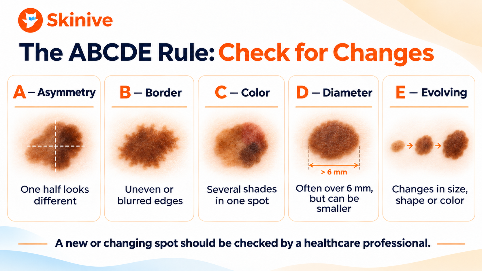
| Sign | What to look for |
|---|---|
| A — Asymmetry | One half of the mole or spot looks different from the other. |
| B — Border | The edges are uneven, irregular, notched or blurred. |
| C — Color | The color is uneven or the spot contains several shades. |
| D — Diameter | A larger spot can be a warning sign, but melanoma can also be smaller than 6 mm. |
| E — Evolving | The spot is changing in size, shape, color, height or appearance. |
Evolving is especially important: a noticeable change deserves attention even if the spot does not meet the other criteria.
The ABCDE rule is a guide, not a diagnosis. Not every melanoma follows the rule, and some benign moles have one or more of these features. The National Cancer Institute’s guide to moles and melanoma explains these signs in more detail.
Look for a Spot That Stands Out
Another useful clue is the “ugly duckling” sign: a mole or spot that looks noticeably different from the others on your skin.
For example, if most of your moles are small and evenly colored but one looks distinctly different, it is worth showing it to a healthcare professional. A spot does not need to match every ABCDE sign to deserve attention.
When Should You See a Dermatologist About a Mole?
Arrange a professional skin examination if you notice:
- A mole or spot that is new or changing
- A mole that looks noticeably different from your other moles
- Changes in size, shape, color, border or texture
- A spot that bleeds, crusts, oozes or does not heal
- Persistent itching, tenderness or pain
- A new or changing spot on a palm, sole or around a nail
- Any skin change that concerns you
A concerning spot should be assessed promptly, even if it is small or painless. Do not wait for it to develop all the ABCDE signs.
Your personal or family history can also affect how often you need professional skin examinations. Ask a dermatologist about a schedule that is appropriate for you, especially if you have many moles, atypical moles or a history of melanoma.
How to Perform a Skin Self-Exam
Regular self-examination helps you become familiar with your skin and notice changes. Many people find that checking their skin about once a month is a practical routine. If a healthcare professional recommends a different schedule, follow their advice.
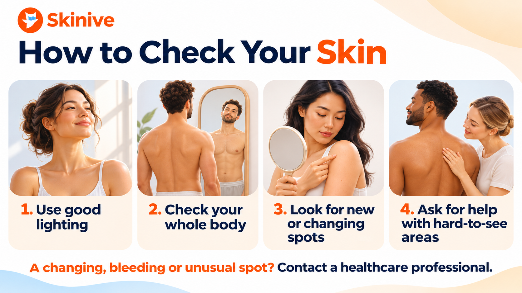
- Choose a well-lit place. Use a full-length mirror and a hand mirror where needed.
- Check your face, ears, neck and scalp. Part your hair or ask someone you trust to help with areas you cannot see.
- Look at your arms, hands and nails. Check between your fingers and on your palms.
- Examine your chest, abdomen, back and sides. Use a mirror or ask for help with your back.
- Check your legs and feet. Include the soles, spaces between the toes and toenails.
- Notice anything new or different. Pay attention to changes in existing spots as well as new ones.
Skin cancer can occur in people of every skin tone and can appear in areas that receive little sun. No matter your skin color, a new or changing spot deserves attention.
If you notice something concerning, contact a healthcare professional rather than waiting until your next planned self-check.
Can You Track Moles with Skinive?
Skinive can help you photograph visible skin concerns and keep a record of previous checks. Saving images of the same area over time can make it easier to describe a change when speaking with a healthcare professional.
For more useful comparisons:
- Take photos in good lighting.
- Keep the area in focus and follow the app’s camera guidance.
- Try to use a similar angle and distance each time.
- Record the location and date of the image.
- Bring your observations and photos to your appointment if you notice a change.
Skinive does not diagnose or rule out melanoma or skin cancer. An app result can be incorrect or inconclusive. If a mole is new, changing, bleeding or otherwise concerning, seek medical advice regardless of the result.
How Does a Dermatologist Examine a Mole?
A dermatologist will usually ask about the spot, how long it has been present and whether it has changed. They may also ask about your personal and family history of skin cancer.
The examination can include dermoscopy: a closer look at the skin using a handheld magnifying device and light. If a spot needs further investigation, the clinician may recommend a biopsy or removal so the tissue can be examined under a microscope.
Not every mole needs to be removed. The decision depends on its appearance, any changes, your history and the clinician’s assessment.
Should You Remove a Mole at Home?
Do not try to cut, burn or remove a mole yourself. Home removal can cause infection or scarring and may make it harder to assess the lesion properly.
If a mole bothers you, is frequently irritated or you would like it removed for cosmetic reasons, talk to a healthcare professional. A suspicious mole should be medically assessed before a removal method is chosen.
How to Protect Your Skin and Moles from the Sun
UV exposure can damage skin and increase the risk of skin cancer. Protecting your skin is useful whether you have a few moles or many.
- Seek shade when the sun is strong.
- Wear protective clothing, a wide-brimmed hat and sunglasses.
- Apply a broad-spectrum sunscreen with SPF 30 or higher to exposed skin.
- Reapply sunscreen regularly, including after swimming, sweating or towel drying.
- Avoid indoor tanning.
- Check the local UV Index when planning time outdoors.
Explore our guide to choosing and applying sunscreen or learn how the Skinive UV Index app can help you follow local UV conditions.
Sunscreen is one part of sun protection. It should be combined with shade and clothing, rather than used as a reason to stay in the sun longer.
Frequently Asked Questions
Are raised moles dangerous?
A raised mole is not automatically dangerous. Many benign moles are raised. However, a raised spot that is new, changing, bleeding or otherwise concerning should be examined by a healthcare professional.
Can a mole appear in adulthood?
Yes. New spots can appear in adulthood, but a new or unusual mole is worth discussing with a healthcare professional, particularly if it looks different from your other moles or changes over time.
Does an atypical mole mean I have melanoma?
No. An atypical mole is not the same as melanoma, and most atypical moles do not become melanoma. However, having several atypical moles is associated with a higher overall risk of melanoma, so ask your dermatologist about appropriate monitoring.
Can melanoma be smaller than 6 mm?
Yes. Some melanomas are smaller than 6 mm. A new, changing or unusual spot should be checked regardless of its size.
Can a mole be pink or skin-colored?
Yes. Moles can be pink, brown, black or close to your natural skin tone. Some melanomas can also have little visible pigment, so color alone cannot establish whether a spot is harmless.
Can a phone app diagnose a mole?
No. An app can help you document and monitor visible skin changes, but it cannot establish a diagnosis or replace a professional skin examination. If a spot concerns you, seek medical advice.
Do all moles need to be removed?
No. Most moles do not need removal. A healthcare professional may recommend removing a mole if it is suspicious, changes, causes repeated irritation or needs further examination.