A halo nevus is a benign mole surrounded by a light or completely depigmented ring of skin. It typically appears as a dark spot or small bump with a “halo” around it. It is not contagious and cannot be passed from person to person.
Halo nevi most commonly develop at a young age, especially in teenagers and people under 25. In most cases, this is a harmless condition that may gradually shrink and even disappear over time.
What to do if you suspect one
If you notice a mole with a white or light ring around it, it’s important not to panic—but also not to ignore it.
The first step is to see a dermatologist. If needed, they may refer you to an oncologist for further evaluation.
Pay attention to:
- the appearance of a new “halo” around a mole
- changes in the size or color of the central area
- itching, pain, or other unusual sensations
- repeated trauma to the lesion
Do not try to remove the mole yourself or use aggressive treatments.
Seeing a doctor helps confirm the nature of the lesion and rule out more serious skin conditions.
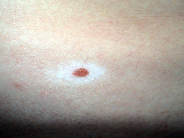
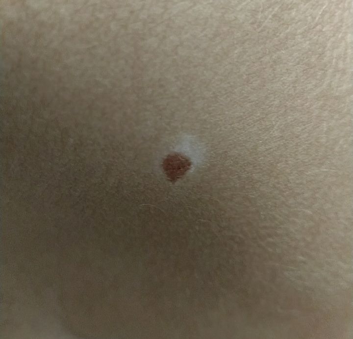
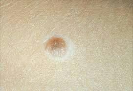
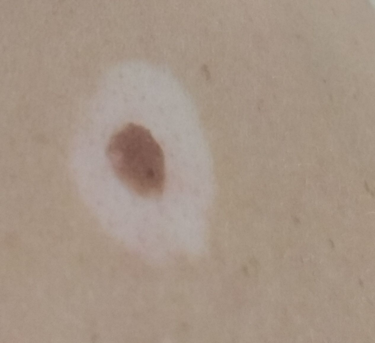
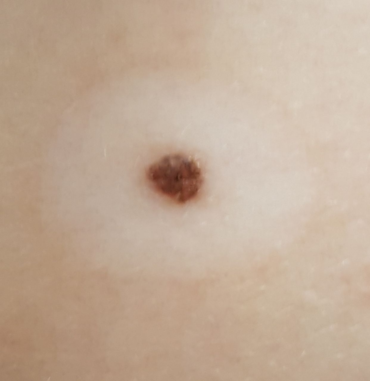
What it looks like
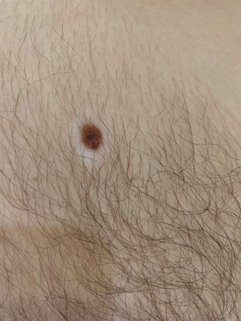
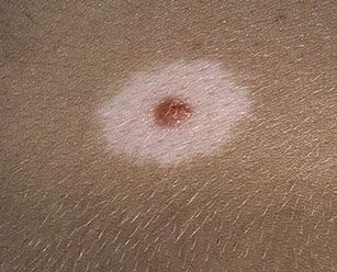
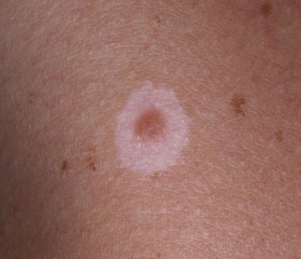
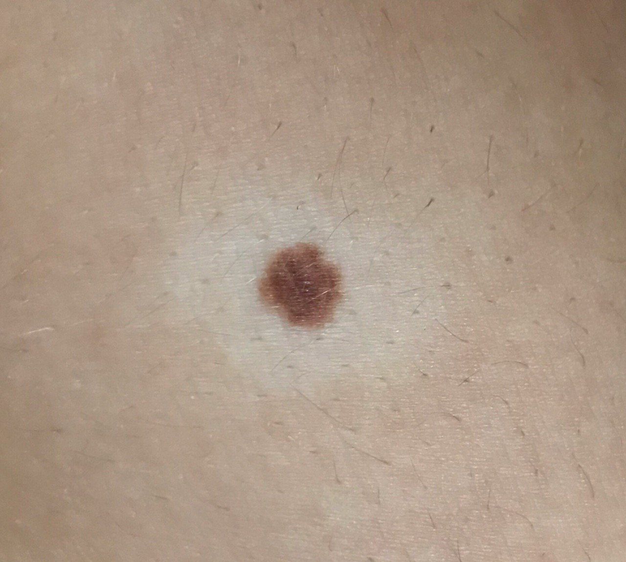
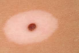
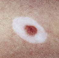
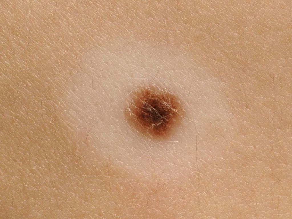
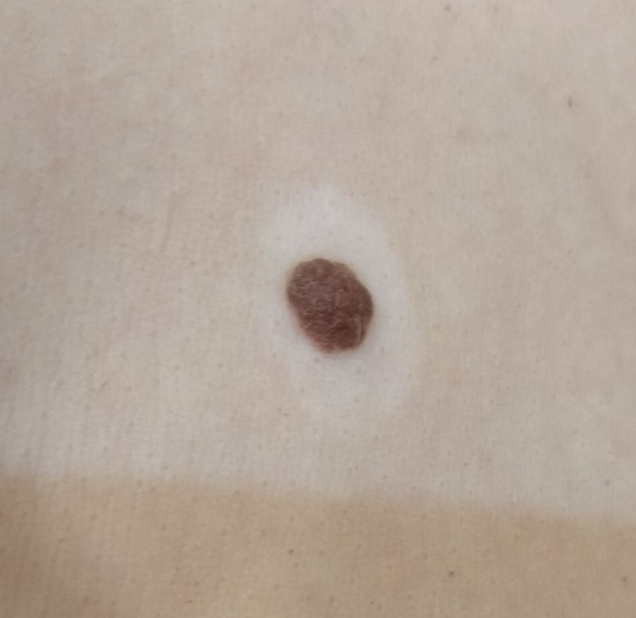
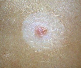
A halo nevus has a characteristic appearance:
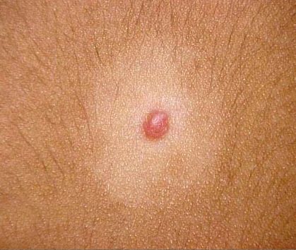
- a small mole in the center (flat or slightly raised)
- a light or white ring surrounding it
The central part is usually:
- round or oval
- with clear borders
- skin-colored to dark brown
The surrounding halo:
- is symmetrical
- may be completely white or slightly pinkish
- often becomes more noticeable after sun exposure
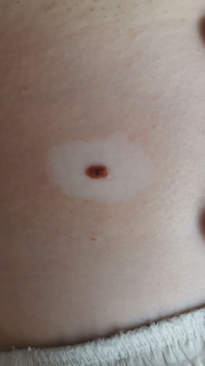
The mole itself is usually up to 1 cm in size, but together with the halo it can reach 3–4 cm.
These lesions most commonly appear on the trunk, less often on other parts of the body.
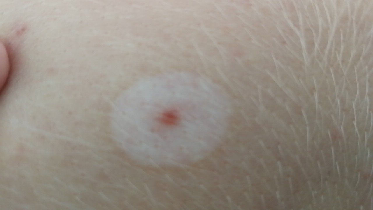
Over time, a halo nevus may change:
- first, the light halo appears
- then the central mole may gradually shrink
- in some cases, the lesion disappears completely
Main symptoms
In most cases, a halo nevus causes no symptoms.
Typically:
- no pain
- no itching
- no discomfort
Occasionally, a few hairs may grow from the center—this is normal.
Any new sensations are a reason to consult a doctor.
Causes
The exact cause of halo nevi is unknown, but several factors may play a role.
Internal factors include:
- genetic predisposition
- the presence of vitiligo (a condition where the skin loses pigment)
- autoimmune processes
An autoimmune reaction occurs when the immune system starts targeting the body’s own cells. In this case, it may destroy pigment-producing cells, leading to the light halo.
External factors include:
- ultraviolet exposure (sunlight, tanning beds)
How it develops
A halo nevus develops in stages.
First, a regular mole appears. Then a light area forms around it because the immune system begins to affect melanocytes (pigment-producing cells), partially destroying them.
Over time, the central mole may shrink or disappear completely. Sometimes only a light patch remains, which may gradually regain its color.
This process usually takes several years.
Types and variations
Halo nevi most often have the classic appearance—a mole with a uniform light halo.
They may vary by:
- size
- how pronounced the halo is
- stage of development (active or regressing)
These differences generally do not affect their safety.
When to see a doctor
You should consult a specialist if:
- the lesion appeared recently and is changing quickly
- the central mole becomes asymmetric
- the color changes or becomes uneven
- itching, pain, or bleeding occurs
It’s also advisable to show a doctor any unusual or suspicious skin changes.
Treatment
In most cases, treatment is not required.
If there are indications (such as diagnostic uncertainty or repeated trauma), surgical removal may be performed, followed by microscopic examination of the tissue.
Key principles:
- monitoring changes
- protecting the skin from sun exposure
- avoiding trauma
The treatment method is always determined by a doctor. Laser or other destructive methods are generally not recommended.
Q&A
Is a halo nevus dangerous?
In most cases, it is harmless and not associated with a high risk of skin cancer.
Can it disappear on its own?
Yes, halo nevi often resolve spontaneously over several years.
Does it need treatment?
Usually not, but monitoring and medical consultation are important.
Why does a white ring appear around the mole?
It’s due to the immune system affecting pigment-producing cells.
Is it related to vitiligo?
Sometimes, as both involve loss of skin pigment.
Can you sunbathe with a halo nevus?
Yes, but skin protection is important since the light area burns more easily.
Should it be removed?
Not if it remains stable and shows no suspicious changes.
How long does it last?
Usually several years, after which it may gradually disappear.
Check Your Skin Instantly
Use the Mole Checker app: Skinive AI to take a photo of a mole or lesion and get an AI-based risk assessment. It helps determine whether professional consultation is recommended, giving you fast guidance and peace of mind.
Sources of medical information
- World Health Organization (WHO)
- American Academy of Dermatology (AAD)
- National Cancer Institute (NCI)
- Fitzpatrick’s Dermatology
- DermNet NZ
🇬🇧 Halo Nevus: When to Seek Medical Advice in the UK
If you notice benign neoplasms, nevi, or moles, it’s important to get a professional opinion. In the UK, you can access dermatology care via the NHS, private clinics, or online consultations.
👉 How to See a Dermatologist in the UK NHS – This main guide explains how NHS referrals work, what to expect from specialist dermatology services, and how to choose between public and private care.
Dermatologists in Major UK Cities:
- Dermatologist in London
- Dermatologist in Manchester
- Dermatologist in Liverpool
- Dermatologist in Birmingham
- Dermatologist in Leeds
- Dermatologists in Other UK cities
Online Dermatology
If you prefer remote care or faster access, try online dermatology consultations. They allow dermatologists to review images, provide advice, and guide next steps without visiting a clinic. Read more in this article: Online Dermatologists in UK.
🇦🇺 Halo Nevus: When to Seek Medical Advice in Australia
If you notice benign neoplasms, nevi, or moles, it’s important to get a professional opinion. In Australia, you can access dermatology care via Medicare (public system), private clinics, or online consultations.
👉 How to See a Dermatologist in Australia – This main guide explains how referrals work through GPs and public clinics, what to expect from specialist dermatology services, and how to choose between public and private care.
Dermatologists in Major Australian Cities:
- Dermatologist in Sydney
- Dermatologist in Melbourne
- Dermatologist in Brisbane
- Dermatologist in Perth
- Dermatologist in Adelaide
- Dermatologists in other Australian cities
Online Dermatology
For faster access or remote care, online dermatology consultations allow dermatologists to review images, provide advice, and guide next steps without visiting a clinic. Read more in this article: Online Dermatologists in Australia.
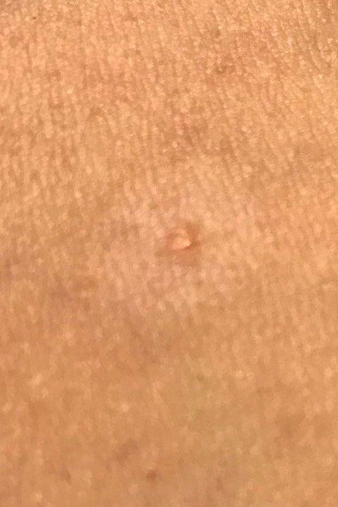
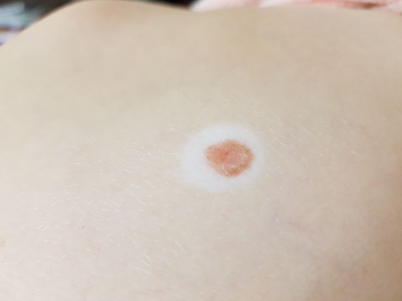
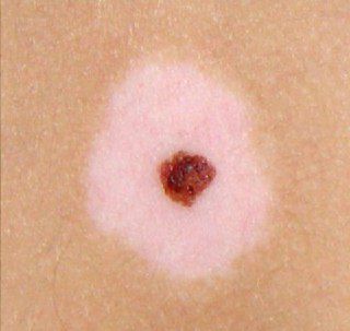
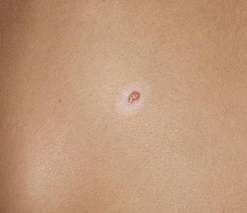
Images of halo nevus:


** Should you identify any copyright infringement regarding the images on this page, kindly reach out to us at info@skinive.com.
Furthermore, please be advised that these photos are not authorized for any purpose.