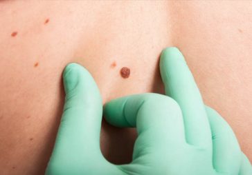
The majority of skin lesions are benign, but when a new lesion or mark appears on our skin, it can be difficult to tell whether it is dangerous. If you have any suspicions about a mark, mole or lesion, you should ask your doctor to check it. Nevertheless, it is useful to know how the common skin lesions look like to be able to recognise them.
Download the Free Skinive App for Skin Scan and use the inbuilt dermatological atlas at any time
In this post, we explain all about the most common skin lesions (with photos) and their main characteristics.
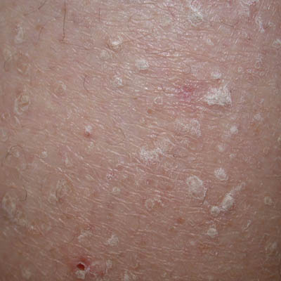
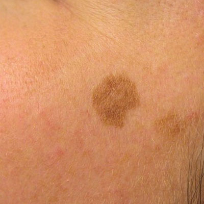
Seborrheic Keratosis
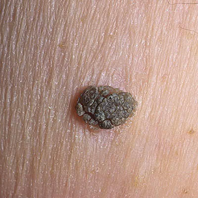
Seborrheic Keratosis, sometimes called senile wart, is a non-cancerous condition that occurs as a light brown, black or tan growth on the surface of the skin. These are usually harmless but may sometimes get irritated or be aesthetically unappealing. They can be removed, if necessary.
Common characteristics:
- These lesions are slightly elevated
- They are waxy and scaly
- They can appear in all areas of the body except for the palms and soles
- They often occur later in life and usually in multiples
- Many times, it has a stuck, “pasted on” appearance
Dermatosis papulosa nigra skin lesion
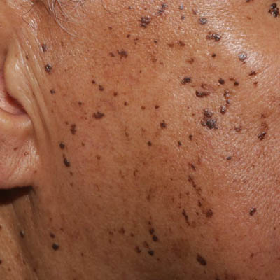
Dermatosis paulosa nigra is a condition that occurs mainly in darker skin types and usually starts to form in adolescence. The lesions are small, darkly pigmented papules that are harmless and generally don’t require treatment.
Common characteristics:
- It appears mainly on cheeks and foreheads
- It often occurs in multiples
- This lesion has a smooth surface
- It appears most likely in an old age
Stucco keratosis skin lesion
Stucco keratosis is another harmless skin condition among common skin lesions with the following characteristics:
· It consists of small, white-gray papules
· It appears mostly on the ankles or feet
· Men are more likely to have it
· It is more common among fair-skinned individuals
Skin tags (acrochordons)
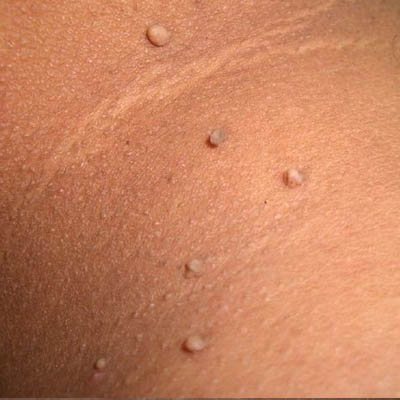
Skin tags or acrochordons are soft skin growths where a narrow papule sticks out of the skin from a short piece of flesh like a tag.
Common characteristics:
- They are fleshy
- They often occur on the eyelids, neck, groin or armpit
- They can become irritated if twisted or rubbed a lot
- They are harmless but can be removed for cosmetic reasons
Cherry angiomas (Hemangioma)
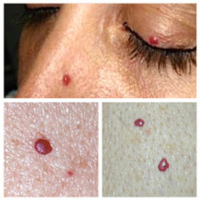
Cherry angiomas are red papules filled with blood vessels made up of capillaries at the surface of the skin.
Common characteristics:
- They usually develop after the age of 40 and increase in number over time.
- They occur in higher concentrations on the trunk of the body.
- They may resemble melanoma when they bleed or clot.
- They do not require treatment, unless for cosmetic reasons.
Dermatofibroma (skin lesion)
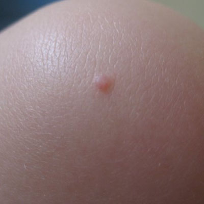
Dermatofibroma is a benign skin tumor that appears as a firm, round, brownish to red-purple growth usually found on the legs.
Common characteristics:
- It feels like a hard lump under the skin
- When squeezed, it dimples since the lesion is tethered to lower layers of the epidermis.
- It initially has a red color, later changing to brown
- It is dome-shaped
- It usually has a darker peripheral rim
- It is often a result of a prior injury
Solar lentigo
Solar lentigos, also known as “sun spots” or “age spots,” are marks on the skin from sun damage that are not cancerous. Although no treatment is required, patients are often at an increased risk for skin cancer and need to exercise precaution.
Sebaceous hyperplasia (skin lesions)
Sebaceous hyperplasia is a skin condition that occurs when sebaceous gland on the skin is enlarged.
Common characteristics:
- It appears as small, skin-colored to yellow papules with a central indent
- These papules often occur on the forehead and central face
- They can often resemble basal cell carcinoma, but they rarely bleed or crust
- Removal is not necessary, unless for cosmetic reasons
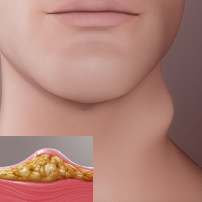
Epidermal inclusion cyst (EIC)
An epidermal inclusion cyst is a common skin cyst, sometimes called sebaceous cyst, even though it arises from hair follicles, not oil glands. The cyst contains a foul-smelling, cheese-like substance formed from degenerating keratinocytes. They can become red and aggravated if the substance enters the dermis, an occurrence which is often mistaken for an infection. Asymptomatic cysts don’t require treatment. Excision can be used to remove a cyst if desired.
Milia or tiny epidermoid cysts
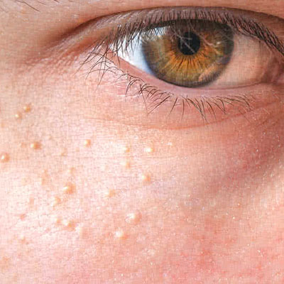
Milia or tiny epidermoid cysts is a condition where small 1-2 mm white to yellow papules occur underneath the surface of the skin.
Common characteristics:
- These cysts can occur in all ages
- They can be extracted without scarring
- They are fixed and long-lasting
- They often occur on the cheeks, eyelids, forehead, and genitalia.
Lipoma
Lipomas are collections of fat under the skin. These common skin lesions are soft and mobile benign tumors that usually stop growing when they reach a few centimeters in diameter. Treatment is surgical and considered elective.
Common characteristics:
- They often appear on the trunk, arms, or thighs
- They tend to occur in multiples in early adulthood
A lipoma is a slow-growing, benign, adipose tumor, often found in the subcutaneous tissues. Lipomas may also occur in deeper tissues such as the intermuscular septa, the abdominal organs, the oral cavity, the internal auditory canal, the cerebellopontine angle and the thorax or any part of the body where there are fat cells. However, they are mostly asymptomatic, unless they press on a nerve causing pain, or rarely develop in the gut wall leading to blockage.
Several factors may increase your risk of developing a lipoma, including:
- Middle age Lipomas are most common in the age group of 40 to 60, and typically rare in children.
- Other disorders People with Dercum’s disease (painful lumps on the trunk, shoulders, arms and legs), Cowden syndrome (hamartomas present in the skin, mucous membranes, thyroid gland, and breast tissue) and Gardner’s syndrome (intestinal polyposis, cysts and osteomas), have an intensified risk of developing multiple lipomas.
- Genetics There is an uncommon autosomal dominant condition called familial multiple lipoma in which groups of fat cells occur under the skin and then produce multiple symmetrical lumps. This runs in families.
Treatment for a lipoma usually isn’t necessarily unless it starts to bother. It is usually carried out in the following ways:
- Surgery Most lipomas are extracted surgically. The side effects are scars and bruises which may possibly remain. Recurrences are not common.
- Liposuction Since lipomas are fat-based, this procedure can work well to reduce its size. A needle and large syringe is used to remove the fatty tissue.
- Steroid injections Steroid injections may also be used right on the affected area. Their use before the surgery is being studied. This treatment shrinks the size of lipoma but usually doesn’t remove it. The reduced lump can then be removed by surgery.
A cancerous, fatty tumor is termed as liposarcoma. Liposarcomas grow rapidly and are usually painful. There is a very little chance that a lipoma turns out to be a malignant liposarcoma and the suspected lump is examined through a biopsy, MRI or CT scan.
Images courtesy of the American Academy of Dermatology