Bowen’s disease is an early form of skin cancer in which abnormal cells are located only in the outermost layer of the skin and have not yet invaded deeper tissues. This is called “carcinoma in situ” (in place).
This is important because at this stage the disease does not metastasize and is usually highly treatable. When detected early, the prognosis is favorable.
It most often appears in people over 35–40 years old. It presents as a slowly growing patch or plaque on the skin. The condition is not contagious and is not transmitted from person to person.
What to do if you suspect it
If you notice an unusual patch or plaque on the skin that does not go away or slowly increases in size, it is important to see a doctor.
The first step is to book an appointment with a dermatologist or oncologist. The specialist will examine the skin, may perform dermoscopy (magnified examination), and may order a biopsy — analysis of a small tissue sample.
Additionally:
- if the skin area is scaly, crusted, or slowly growing, do not delay a medical visit
- if findings are unclear, the doctor may suggest monitoring with regular follow-ups
It is important not to attempt self-treatment or removal, as this may complicate diagnosis and increase the risk of complications.
Early consultation allows detection at a stage where treatment is most effective and least invasive.
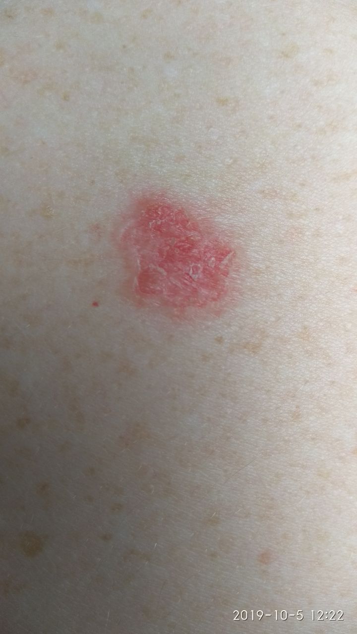
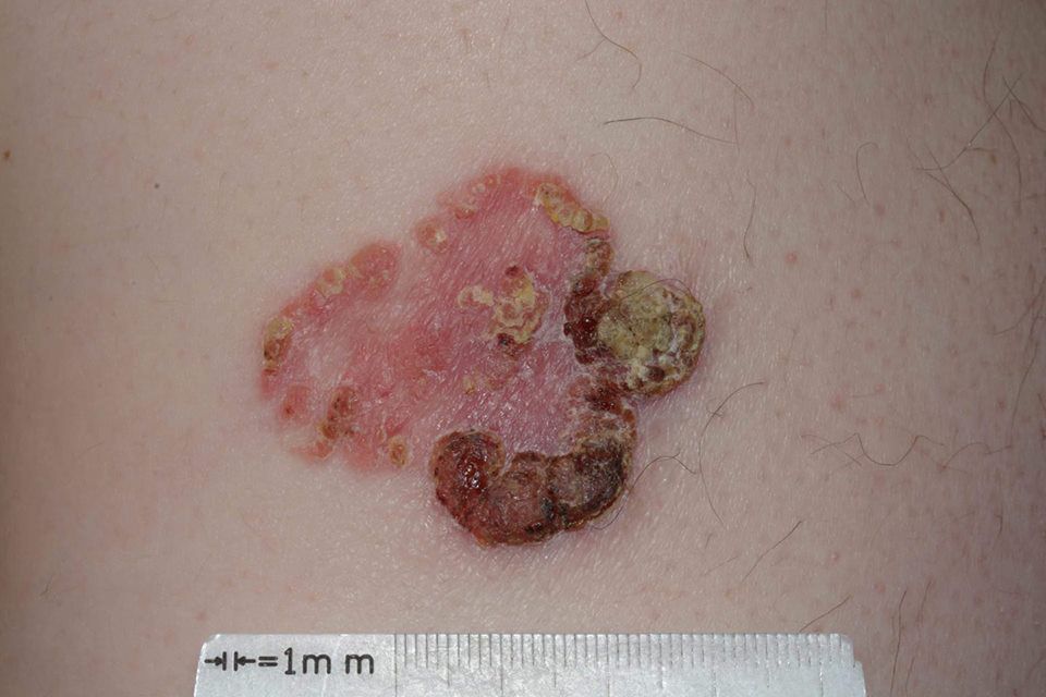
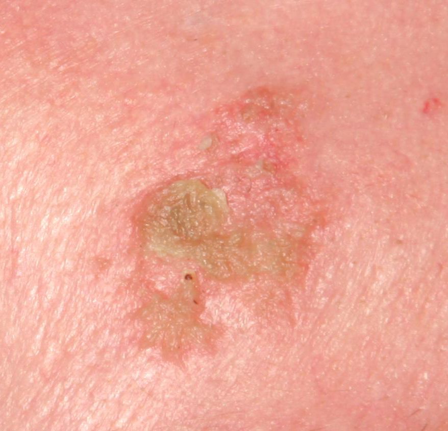
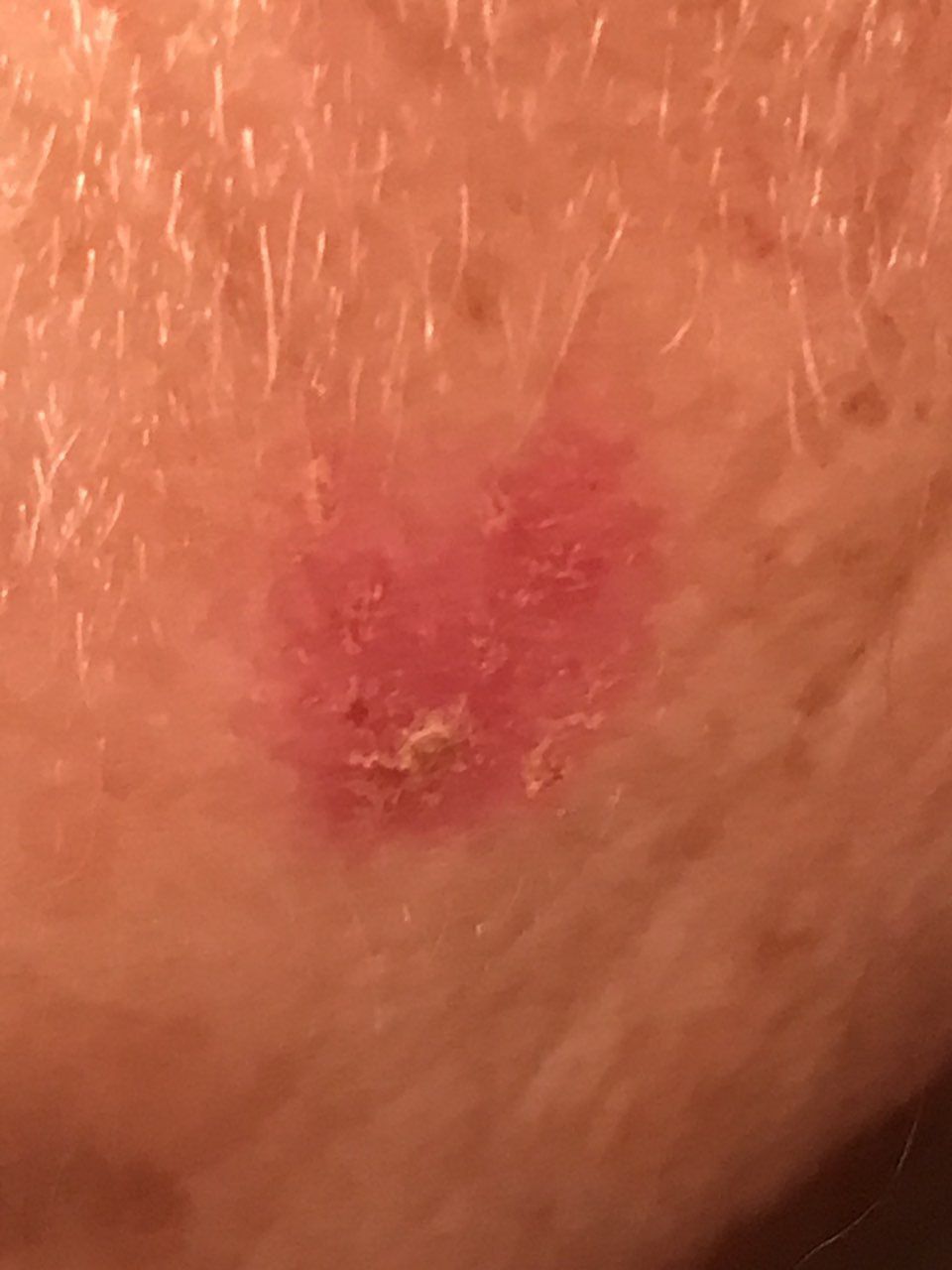
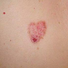
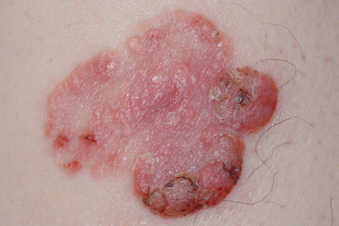
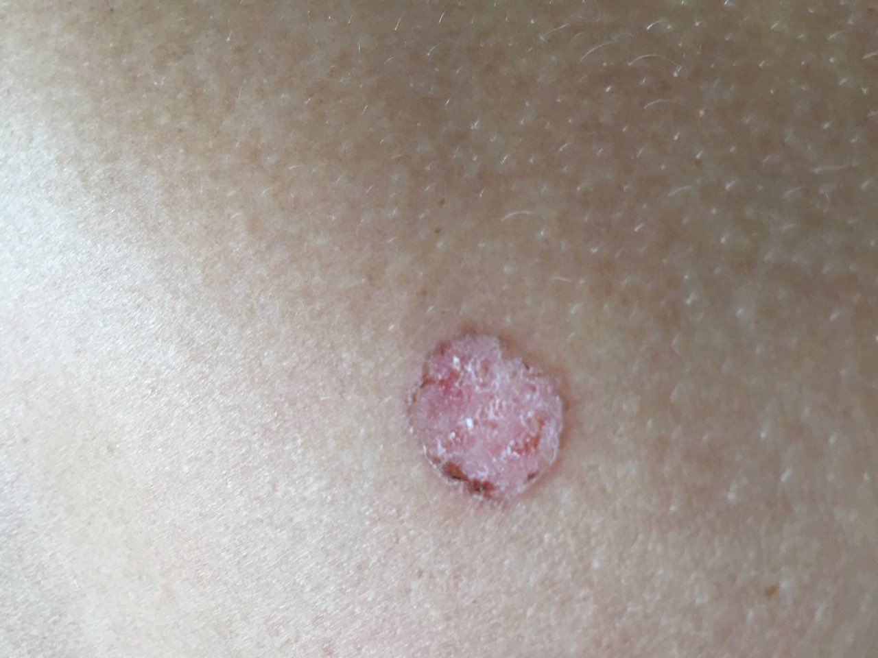
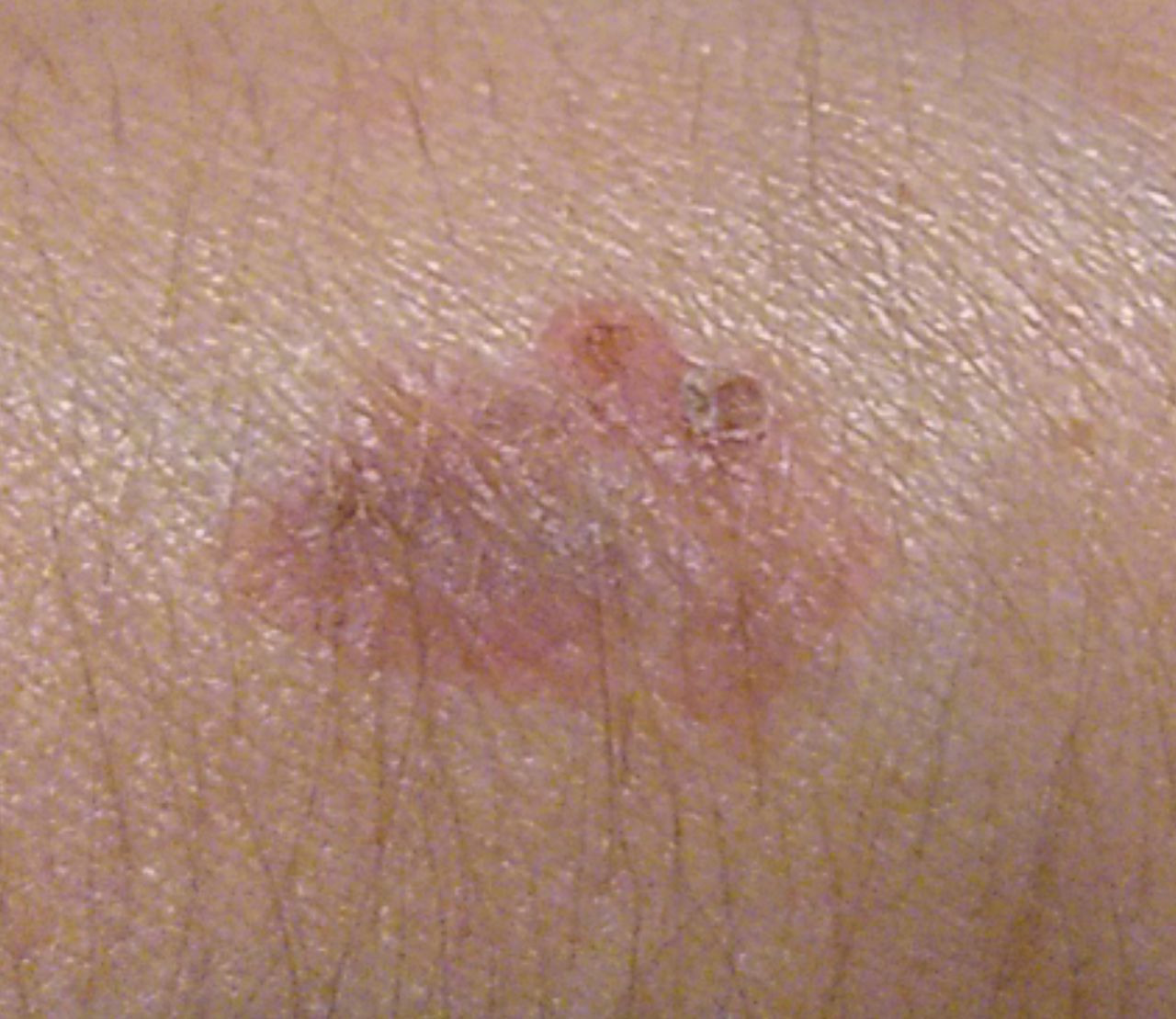
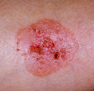
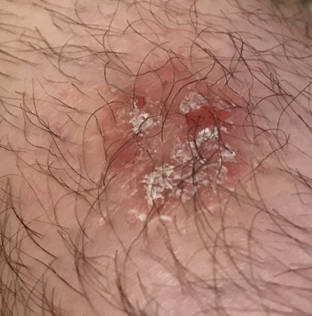
What the disease looks like
Bowen’s disease usually appears as a patch or plaque on the skin.
Characteristic features:
- pink or reddish color
- well-defined but irregular borders
- rough, dry surface
The surface may be:
- covered with crusts
- showing areas of scaling
- sometimes with small erosions (superficial skin damage)
The lesion is usually only slightly raised and grows very slowly — sometimes over years.
It most commonly appears on the trunk, face, neck, hands, or scalp.
Below in the article you can view real photos to better understand how the condition looks.
Main symptoms
In early stages, the disease often causes no noticeable discomfort.
Possible signs include:
- a long-lasting patch or plaque
- slow enlargement of the lesion
- scaling and dryness
- formation of crusts
Sometimes mild itching or burning may occur, but significant pain is usually absent.
Causes of the disease
The exact cause of Bowen’s disease is unknown, but several risk factors increase the likelihood of developing it.
External factors:
- long-term exposure to ultraviolet radiation (sun, tanning beds)
- ionizing radiation
- contact with skin-damaging substances
- chronic skin trauma
Internal and additional factors:
- certain chronic skin conditions
- possible role of human papillomavirus (HPV)
- age
How the disease develops
Bowen’s disease develops in the outer layer of the skin (epidermis).
In simple terms, skin cells begin to divide abnormally but remain on the surface and do not invade deeper layers. This is why the condition is considered an early form of cancer.
Over time, without treatment, these cells may invade deeper layers of the skin, progressing into invasive cancer.
Forms and types
Bowen’s disease may appear in different ways: as a single patch or multiple lesions.
They may vary in size, color, and degree of scaling. Despite these differences, the underlying condition remains the same — a superficial skin malignancy.
When to see a doctor
You should consult a specialist if:
- a patch or plaque persists for a long time
- the lesion gradually enlarges
- scaling, crusting, or erosions appear
- any changes occur in an existing lesion
It is also recommended to regularly examine the skin, especially with frequent sun exposure.
Treatment
The main treatment is removal of the affected skin area. Surgical excision is most commonly used, where the lesion is completely removed.
In some cases, radiation therapy may be used.
The treatment method is selected individually by the doctor based on size, location, and characteristics of the lesion.
Additionally, it is important to:
- protect the skin from sun exposure
- avoid trauma to the area
- attend regular follow-up examinations
Self-treatment is not recommended, as it may increase the risk of recurrence or progression.
Q&A
Can Bowen’s disease be cured?
Yes, in early stages it is usually successfully treated with appropriate management.
How dangerous is it?
At an early stage the risk of spread is minimal, but without treatment it may progress.
How is Bowen’s disease treated?
The main method is removal of the lesion. The specific approach is chosen by a doctor.
Is it already cancer or not?
It is an early form of skin cancer in which cells have not yet invaded deeper layers.
Can it go away on its own?
No, without treatment the lesion usually persists and slowly enlarges.
Are there risks of complications?
Yes, without treatment it may progress to a more serious form of skin cancer.
Can it be prevented?
Reducing sun exposure and regular skin checks can lower the risk.
Is follow-up needed after treatment?
Yes, regular check-ups help detect any new changes early.
Check Your Skin Instantly
Use the Skin cancer detector: Skinive AI to take a photo of a mole or lesion and get an AI-based risk assessment. It helps determine whether professional consultation is recommended, giving you fast guidance and peace of mind.
Sources
- World Health Organization (WHO)
- National Cancer Institute (NCI)
- American Academy of Dermatology (AAD)
- NCCN Clinical Practice Guidelines in Oncology (Squamous Cell Skin Cancer)
🇬🇧 Bowen’s disease: How Diagnosis and Treatment Work in the UK
If you notice suspicious or skin cancer lesions, such as a new growth, persistent sore, or changes in an existing spot—including a raised bump, ulceration, or slow-healing lesion—it’s important to contact your GP immediately and seek professional care, which in the UK is available via the NHS, private clinics, or online dermatology consultations.
👉 How to See a Dermatologist in the UK NHS – This main guide explains how NHS referrals work, what to expect from specialist dermatology services, and how to choose between public and private care.
Dermatologists in Major UK Cities:
- Dermatologist in London
- Dermatologist in Manchester
- Dermatologist in Liverpool
- Dermatologist in Birmingham
- Dermatologist in Leeds
- Dermatologists in Other UK cities
Online Dermatology
If you prefer remote care or faster access, try online dermatology consultations. They allow dermatologists to review images, provide advice, and guide next steps without visiting a clinic. Read more in this article: Online Dermatologists in UK.
🇦🇺 Bowen’s disease: How Diagnosis and Treatment Work in Australia
If you notice suspicious or skin cancer lesions, such as a new growth, persistent sore, or changes in an existing spot—including a raised bump, ulceration, or slow-healing lesion—it’s important to contact your GP immediately and seek professional care. In Australia, dermatology care is available via Medicare (public system), private clinics, or online consultations.
👉 How to See a Dermatologist in Australia – This main guide explains how referrals work through GPs and public clinics, what to expect from specialist dermatology services, and how to choose between public and private care.
Dermatologists in Major Australian Cities:
- Dermatologist in Sydney
- Dermatologist in Melbourne
- Dermatologist in Brisbane
- Dermatologist in Perth
- Dermatologist in Adelaide
- Dermatologists in other Australian cities
Online Dermatology
For faster access or remote care, online dermatology consultations allow dermatologists to review images, provide advice, and guide next steps without visiting a clinic. Read more in this article: Online Dermatologists in Australia.
Images of Bowen disease:


** Should you identify any copyright infringement regarding the images on this page, kindly reach out to us at info@skinive.com.
Furthermore, please be advised that these photos are not authorized for any purpose.